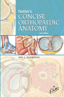
About The Book
Introducing the new and definitive orthopedic anatomy atlas for students and clinicians! This concise, easy-to-use atlas of orthopedic anatomy uses Dr. Frank Netter images from both the Atlas of Human Anatomy and the 13-volume Netter Collection of Medical Illustrations.
- Tables listing key information on bones, joints, muscles, and nerves highlight each Netter image.
- Each chapter contains useful clinical information on disorders, trauma, history and physical exam, radiology, surgical approaches, and minor procedures.
- Information is presented in tabular format for easy reference.
- Key material is highlighted in different colors depending on its content.
- 450 Netter images in a portable 6″ x 9″ format.
- Four-color design throughout.


Add Comment